News Releases
Konica Minolta Launches Joint Research with Institut Pasteur and BioAxial in Developing Support System for Pharmaceutical Development
- Leading-edge bioimaging technologies to speed process of new drug development -
Tokyo (June 6, 2016) - Konica Minolta, Inc. (Konica Minolta), a global technology company with distinctive core competence in chemistry and imaging, has launched with the Institut Pasteur and Paris-based BioAxial a joint research project regarding bioimaging technologies to support pharmaceutical development.
Joint Research
The joint research aims to develop in vivo fluorescent nanoparticles and an observation system. The system will allow direct observation of the movement and distribution of molecules or cells within mice’s body and further observation of effects of the drug on live cells when delivered to organs and cells (living cell imaging). These functions are expected to enable observation of effect of drugs and mechanism of actions and support accurate evaluation of the effectiveness of new drug.
Konica Minolta is willing to utilize its development of proprietary fluorescent nanoparticles and analyzing technologies of their images through technological cooperation with the Institut Pasteur’s imaging technologies necessary to meet the demands of pharmaceutical development and BioAxial’s microscopic observation device which can provide super-resolved images. The joint research is expected to develop and offer new in vivo* imaging technologies.
*in vivo (defined as “within living organism”): test articles are directly administered to laboratory animals for detecting reactions of the drug in the living organs and cells
Konica Minolta’s Proprietary Technologies
One of hardships in the development of new drugs has been the low success rate of the candidate medicine’s efficacy at the clinical study. To grapple with the challenge, it is effective to use detailed analysis of the medicine’s effectiveness within cells based on quantification of protein, among others, when researchers study candidate medicines. Such detailed analysis requires innovative imaging technologies. In addition, detection technologies with the use of fluorescent dyes is a field of fluorescent detection technologies for research and development of cell imaging and bioimaging. Conventional marker techniques using fluorescent dyes are subject to problems such as being photobleaching, low sensitivity and low quantitative capability.
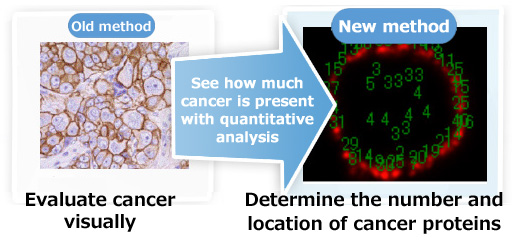
Konica Minolta addressed these problems by applying techniques used in its own silver halide particle development technologies fostered for photographic films. The company has successfully developed fluorescent nanoparticles that are both approximately 30,000 times as bright as conventional fluorescent dyes and highly photostable. In addition, Konica Minolta has developed “nanoparticle surface modification technique” to create better compatibility to biological materials, including bonding of the fluorescent nanoparticle and antibody, and “fluorescent bright spot analysis software” so that the quantitative capability of diagnosis is improved, based on “single particle fluorescent imaging” that can count protein in the cells and cancer tissues by single particle.

Prospects for Business Development
Konica Minolta launched a “pathology specimen production” program in the in-vitro field in Japan in July 2015, utilizing its fluorescent nanoparticle technologies, and has been highly valued by many pharmaceutical companies and drug discovery-related companies. While driving the business development of in-vitro diagnostic services, Konica Minolta expects to enter in a new business for consignment of drug development from pharmaceutical companies, based on the fundamental technologies in in-vivo imaging from the joint research. As part of the joint research, pharmaceutical companies will be able to consign Konica Minolta to apply their candidate drugs to cells and animals and conduct nano-scale observation so that the effectiveness and side effects be estimated and screening be made. Globally many pharmaceutical companies and drug discovery-related companies have shown interest in the prospects of such technologies, as expectation has been growing for improvement in the success rate of clinical tests in the future. Furthermore, a new business development into light imaging contrast business for visualized diagnosis of cancer and other illness during clinical tests and diagnosis, replacing the existing PET (positron emission tomography) imaging.
The joint research has come to the stage of launch, as Konica Minolta’s advanced material technologies regarding the new fluorescent nanoparticle imaging have been highly recognized by the Institut Pasteur and BioAxial, two French institutions renowned for their state-of-the-art research.
Under the brand proposition “Giving Shape to Ideas,” Konica Minolta aims to contribute to creating social values and become a company vital to society, by delivering new services and values.
- Part of this research has been assigned by the New Energy and Industrial Technology Development Organization (NEDO) through its “Japan-France Bilateral R&D Cooperation Program.”
